Video based object tracking
| |
| Research Taxonomy |
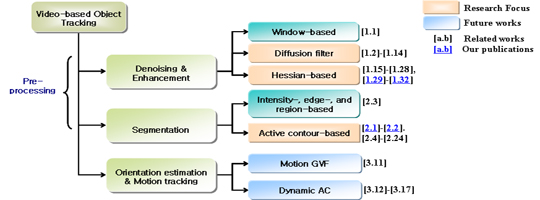 |
| Research Content |
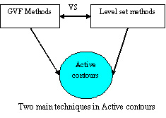
Active contours -- or snakes -- are computer-generated curves that move within images to find object boundaries (note that the 3D version is often known as deformable models or active surfaces in the literature). They are often used in computer vision and image analysis to detect and locate objects, and to describe their shape.
Applications:
- Tracking identified person(s)/object(s) in smart service systems (CAMUS), films making, control systems (robotics).
- In biomedical images: tracking leukocytes, stem cells etc.
Our team has been doing research on motion tracking problems which usually have three following steps: |
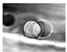
1. Target segmentation using active contours (snakes) |
|
The U-shaped object and the snake evolving to its boundary |
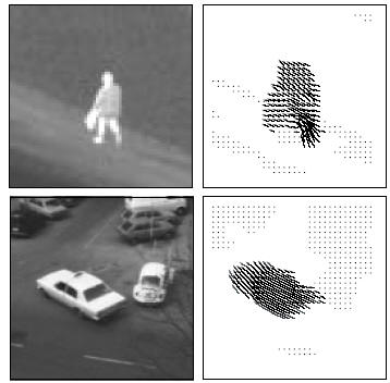
| 2. Motion field estimation |
| Tensor based technique |
|
| 3. Motion tracking |
| Motion GVF techniques |
|
The moving leukocyte and its motion tracking |
| References |
1. Denoising & enhancement
[1.1] R.C. Gonzalez and R.E. Woods, “Digital Image Processing”, Prentice Hall, New Jersey, U.S., 2002
[1.2] P. Perona and J. Malik, “Scale space and edge detection using anisotropic diffusion”, IEEE Trans. Pattern Anal. Mach. Intell., vol 12, pp. 629-636, 1990.
[1.3] P. Perona, T. Shiota, and J. Malik, “Anisotropic diffusion”, B.M. ter Haar Romeny (Ed), Geometry driven diffusion in computer vision, Kluwer, pp. 72-92, 1994.
[1.4] J. Weikert, “A Review of Nonlinear Diffusion Filtering”, Scale Space Theory in Computer Vision, LNCS, vol. 1252, pp. 3-28, 1997.
[1.5] P. Perona, “Orientation Diffusions”, IEEE Trans. Image Processing, vol. 7, no. 3, March 1998.
[1.6] B. Tang, G. Sapiro, and V. Caselles, “Direction Diffusion”, IEEE Int. Conf. on Computer Vision, vol 2, 1245-1252, 1999.
[1.7] O. Scherzer and J. Weikert, “Relations Between Regularization and Diffusion Filtering”, Journal of Mathematical Imaging and Vision ,vol. 12, pp. 43–63, 2000.
[1.8] J. Suri, J. Gao, S. Singh, and S. Laxminarayan, “A Comparison of State-of-the-Art Diffusion Imaging Techniques for Smoothing Medical/Non-Medical Image Data”, IEEE Int. Conf. on Pattern Recognition, vol. 1, pp. 508- 511, 2002.
[1.9] H. Scharr and J. Weickert, “An Anisotropic Diffusion Algorithm with Optimized Rotation Invariance”, in G. Sommer, N. Kruger, C. Perwass (Eds.), Mustererkennung 2000.
[1.10] J. Weickert and H. Scharr, “A Scheme for Coherence-Enhancing Diffusion Filtering with Optimized Rotation Invariance”, CVGPR Group Technical Report at the Department of Mathematics and Computer Science, University of Mannheim, Germany, TR 4/2000.
[1.11] Y. Yu and S. Acton, “Speckle Reducing Anisotropic Diffusion”, IEEE Trans. Image Processing, vol. 11, no. 11, November 2002.
[1.12] H. Yu and C. Chua, “GVF-Based Anisotropic Diffusion Models”, IEEE Trans. Image Processing, vol. 15, no. 6, June 2006.
[1.13] H. Luo, L. Zhu, and H. Ding, “Coupled anisotropic diffusion for image selective smoothing”, Signal Processing, vol. 86, pp. 1728–1736, 2006.
[1.14] M. M. Orkisz, “Improved vessel visualization in MR angiography by nonlinear anisotropic filtering”, MRM, vol. 37, no. 6, pp. 914- 919, June 1997.
[1.15] A.F. Frangi, W.J. Niessen, K.L. Vincken, and M.A. Viergever, “Multiscale vessel enhancement filtering”, Proc. Int. Conf. Medical Image Computing Computer-Assisted Intervention, LNCS, vol. 1496, pp. 130-137, 1998.
[1.16] Y. Sato, S. Nakajima, N. Shiraga, H. Atsumi, S. Yoshida, T. Koller, G. Gerig, and R. Kikinis, “Three-dimensional multi-scale line filter for segmentation and visualization of curvilinear structures in medical images”, Medical Image Analysis, vol. 2, no. 2, pp. 143-168, 1998.
[1.17] C. Lorenz, I.-C. Carlsen, T.M. Buzug, C. Fassnacht, and J. Weese, “A multi-scale line filter with automatic scale selection based on the Hessian matrix for medical image segmentation”, in Proc. Scale-Space Theories in Computer Vision, LNCS, 1997, vol. 1252, pp.152-163.
[1.18] K. Krissian, G. Malandain, and N. Ayache, “Model based detection of tubular structures in 3D images”, Tech. Rep., INRIA 373, France, 1999.
[1.19] K. Krissian, G. Malandain, and N. Ayache, “Directional anisotropic diffusion applied to segmentation of vessels in 3D images”, in Proc. Int. Conf. Scale-Space, 1997, pp. 345-348.
[1.20] K. Krissian, G. Malandain, N. Ayache, R. Vaillant, and Y. Trousset, “Model based multiscale detection of 3D vessels”, in Proc. IEEE Int. Conf. Computer Vision Pattern Recognition, 1998, pp. 722-727.
[1.21] H. Shikata, E. A. Hoffman, and M. Sonka, “Automated segmentation of pulmonary vascular tree from 3D CT images”, in Proc. SPIE Int. Symp. Medical Imaging, San Diego, CA, 2004.
[1.22] G. Agam, III S.G. Armato, and C. Wu, “Vessel tree reconstruction in thoracic CT scans with application to nodule detection”, IEEE Trans. Med. Imag., vol. 24, no. 4, pp. 486-499, Apr. 2005.
[1.23] G.H. Bamberger and M. J. T. Smith, “A filter bank for the directional decomposition of images - theory and design”, IEEE Trans. Sig. Proc., vol. 40, pp. 882-893, 1992.
[1.24] S. Park, M. J. T. Smith, and J. J. Lee, “Fingerprint enhancement based on the directional filter bank”, in IEEE Int. Conf. Image Proc. (ICIP'00), 2000, pp. 793-796.
[1.25] S. Park, M. J. T. Smith, and R. M. Mersereau, “A new directional filter bank for image analysis and classification”, ICASSP, vol. 3, pp. 1417-1420, 1999.
[1.26] S. Park, M. J. T. Smith, and R. M. Mersereau, “Improved structures of maximally decimated directional filter banks for spatial image analysis”, IEEE Trans. Image Processing, vol. 13, no. 11, pp. 1424-1431, Nov. 2004.
[1.27] R. H. Bamberger, “The directional filter bank: A multirate filterbank for the directional decomposition of images”, Ph.D. thesis, Georgia Tech., Nov. 1990.
[1.28] R. H. Bamberger and M. J. T. Smith, “A multirate filter bank based approach to the detection and enhancement of linear features in images”, in IEEE. Int. Conf. on Acoustics, Speech and Signal Processing (ICASSP'91), Canada, Apr. 1991, vol. 4, pp. 2557-2560.
[1.29] P. T. H. Truc, Md. A. U. Khan, Y.-K. Lee, S. Y. Lee, and T.-S. Kim, “Vessel Enhancement Filter using Directional Filter Bank”, Computer Vision and Image Understanding, in press, 2008.
[1.30] Md. A. U. Khan, P. T. H. Truc, R. Bahadur, and S. Javed, “Vessel Enhancement using Directional Features”, Information Technology Journal 6(6), pp. 851-857, 2007.
[1.31] P. T. H. Truc, Md. A. U. Khan, S. Y. Lee, and T.-S. Kim, "A New Approach to Vessel Enhancement in Angiography Images", IEEE/ICME Int. Conf. on Complex Med. Eng., pp. 878-884, Beijing, China, May 23-27, 2007.
[1.32] P. T. H. Truc, Md. A. U. Khan, S. Y. Lee, and T. S. Kim, “Vessel Enhancement using Decimation-free Directional Filter Bank”, US Patent (application number: 11/892,030). |
2. Segmentation
[2.1] P. T. H. Truc, Y. H. Kim, Y.-K. Lee, S. Y. Lee, and T.-S. Kim, “Evaluation of Active contour-based techniques toward Bone segmentation for CT images”, in Springer IFMBE Proceedings, vol 14, pp. 2997-3000, 2006.
[2.2] P. T. H. Truc, T.-S. Kim, Y.-K. Lee, and S. Y. Lee “Automatic Bone Segmentation from CT Images using Chan-Vese Multiphase Active Contour”, J. Biomed. Eng. Res., vol 28, no. 6, pp. 713-720, 2007.
[2.3] L. Wang, M. Greenspan, and R. Ellis, “Validation of bone segmentation and improved 3-D registration using contour coherence in CT data”, IEEE Trans. Med. Imag., vol. 25, pp. 324-334, 2006.
[2.4] M. Kass, A. Witkin, and D. Terzopoulos, “Snakes: active contour models”, Int. J. Comput. Vis., vol. 1, no. 4, pp. 321-331, 1988.
[2.5] L. Cohen and I. Cohen, “Finite element methods for active contour models and balloons for 2D and 3D images”, IEEE Trans. Patt. Anal. Mach. Intell., vol. 15, no. 11, pp. 1131-1147, 1993.
[2.6] C. Xu and J. Prince, “Snakes, shapes, and gradient vector flow”, IEEE Trans. Image Proc., vol. 7, pp. 359-369, 1998.
[2.7] Caselles, F. Catte, T. Coll, and F. Dibos, “A geometric model for active contours”, Numerische Mathematik, vol. 66, pp. 1-31, 1993.
[2.8] V. Caselles, R. Kimmel, and G. Sapiro, “Geodesic active contours”, Int. J. Comp. Vis., vol. 22, pp. 61-79, 1997.
[2.9] N. Paragios, O.M-Gottardo, and V. Ramesh, “Gradient vector flow fast geometric active contours”, IEEE Trans. Patt. Anal. Mach. Intell., vol. 26, pp. 402-407, 2004.
[2.10] L. Vese and T. Chan, “A multiphase level set framework for image segmentation using Mumford and Shah model”, Int. J. Comp. Vis., vol. 50, no. 3, pp. 271-293, 2002.
[2.11] J. Sethian, “Level set methods = evolving interface in geometry, computer vision”, New York, USA: Cambridge University Press, 1996.
[2.12] S. Osher and R. P. Fedkiw, “Level set methods and dynamic implicit surfaces”, New York, USA: Springer-Verlag, 2003.
[2.13] T. Sebastian, H. Tek, J. Crisco, and B. Kimia, “Segmentation of carpal bones from CT images using skeletally coupled deformable models”, Medical Image Analysis, vol. 7, pp. 21-45, 2003.
[2.14] C. S. Poon and M. Braun, “Image segmentation by a deformable contour model incorporating region analysis”, Phys. Med. Biol., vol. 42, pp. 1833-1841, 1997.
[2.15] X. M. Pardo, M. J. Carreira, A. Mosquera, and D. Cabello, “A snake for CT image segmentation integrating region and edge information”, Image Vis. Comput., vol. 19, pp. 461-475, 2001.
[2.16] H. Tek and B. Kimia, “Volumetric segmentation of medical images by three-dimensional bubbles”, Computer Vision and Image Understanding, vol. 64, pp. 246-258, 1997.
[2.17] S. Zhu and A. Yuille, “Region competition: unifying snakes, region growing, and Bayes/MDL for multiband image segmentation”, IEEE Trans. Patt. Anal. Mach. Intell., vol. 18, pp. 884-900, 1996.
[2.18] R. Malladi, J. A. Sethian, and B. C. Vemuri, “Shape modeling with front propagation: a level set approach”, IEEE Trans. Patt. Anal. Mach. Intell., vol. 17, no. 2, pp. 158-175, 1995.
[2.19] L. Ballerini and L. Bocchi, “Multiple genetic snakes for bone segmentation”, in Applications of Evolutionary Computing: EvoWorkshops, ser. LNCS, vol. 2611, 2003, pp. 346-356.
[2.20] S. Osher and J. A. Sethian, “Fronts propagating with curvature dependent speed: algorithms based on Hamilton-Jacobi formulations”, J. Comp. Physics, vol. 79, pp. 12-49, 1998.
[2.21] V. Caselles, R. Kimmel, and G. Sapiro, “Geodesic active contours”, in Int. Conf. Comp. Vis., 1995, pp. 694.699.
[2.22] A. Yezzi, S. Kichenassamy, A. Kumar, P. Olver, and A. Tannenbaum, “A geometric snake model for segmentation of medical imagery”, IEEE Trans. Med. Imag., vol. 16, pp. 199-209, 1997.
[2.23] T. Chan and L. Vese, “Active contours without edges”, IEEE Trans. Image Proc., vol. 10, pp. 266-277, 2001.
[2.24] D. Mumford and J. Shah, “Optimal approximation by piecewise smooth functions and associated variational problems”, Commun. Pure Appl. Math., vol. 42, pp. 577-685, 1989. |
3. Visual tracking
[3.1] G. Welch and E. Foxlin, “Motion tracking: No silver bullet, but a respectable arsenal,” IEEE Comput. Graph. Appl., vol. 22, no. 6, pp. 24–38, 2002.
[3.2] S. Julier and G. Bishop, “Tracking: How hard can it be?,” IEEE Comput. Graph. Appl., vol. 22, no. 6, pp. 22–23, 2002.
[3.3] D. J. Kriegman, G. D. Hager, and A. S. Morse, Eds.,” The Confluence of Vision and Control”, New York: Springer-Verlag, 1998, vol. 237, Lecture Notes in Control and Information Sciences.
[3.4] I. J. Cox, “A review of statistical data association techniques for motion correspondence,” Int. J. Comput. Vision, vol. 10, no. 1, pp. 53–66, 1993.
[3.5] A. Mitiche and P. Bouthemy, “Computation and analysis of image motion: A synopsis of current problems and methods,” Int. J. Comput. Vision, vol. 19,pp. 29–55, 1996.
[3.6] M. J. Swain and M. A. Stricker, “Promising directions in active vision,” Int. J. Comput. Vision, vol. 12, no. 2, pp. 109–126, 1993.
[3.7] C. Kublbeck and A. Ernst, “Face detection and tracking in video sequences using the modified census transformation”, Image and Vision Computing, vol. 24, pp. 564–572, 2006.
[3.8] Y. Hsiao, C. Chuang, Y. Lu, and J. Jiang, “Robust multiple objects tracking using image segmentation and trajectory estimation scheme in video frames”, Image Vis. Comput., 2006.
[3.9] D. Jackson, J. Yezzi, and S. Soatto, “Tracking Deformable Moving Objects Under Severe Occlusions”, IEEE Conf. on Decision and Control, vol.3, pp. 2990- 2995, 2004.
[3.10] C. Kambhamettua, D. Goldgofb, M. Hec, and P. Laskov, “3D nonrigid motion analysis under small deformations”, Image Vis. Comput., vol. 21, pp. 229–245, 2003.
[3.11] T. Lam and R. Lee, “Visual Tracking by Using Kalman Gradient Vector Flow (KGVF) Snakes”, Proceedings of International Conference Knowledge-Based Intelligent Information and Engineering Systems, pp. 557-563, September 2004.
[3.12] A. Blake and M. Isard, “Active Contours”, Springer Verlag, 1998.
[3.13] I. Dydenko, F. Jamal, O. Bernard, J. Dfihooge, I. E. Magnin, and D. Friboulet, “A level set framework with a shape and motion prior for segmentation and region tracking in echocardiography”, Medical Image Analysis, vol. 10, pp. 162–177, 2006.
[3.14] S. Dambreville, Y. Rathi, and A. Tannenbaum, “Tracking Deformable Objects with Unscented Kalman Filtering and Geometric Active Contours”, Proceedings of the 2006 American Control Conference Minneapolis, Minnesota, USA, June, 2006.
[3.15] M. Niethammer, A. Tannenbaum, and S. Angenent, “Dynamic Active Contours for Visual Tracking”, IEEE Trans. Automatic Control, vol. 51, no. 4, April 2006.
[3.16] N. Paragios and R. Deriche, “Geodesic Active Contours and Level Sets for the Detection and Tracking of Moving Objects”, IEEE Trans. Patt. Anal. Mach. Intell., vol. 22, no. 3, March 2000.
[3.17] Y. Rathi, N. Vaswani, A. Tannenbaum, and A. Yezzi, “Particle Filtering for Geometric Active Contours with Application to Tracking Moving and Deforming Objects”, IEEE Conf. on CVPR, vol. 2, pp. 2-9, 2005. |
|
|
| :: UC Lab News |
06-24-2008
New website launched by Activity Recognition team...
|
|